
Development of a Class Prediction Model to Discriminate Pancreatic Ductal Adenocarcinoma from Pancreatic Neuroendocrine Tumor by MALDI Mass Spectrometry Imaging
Highlights
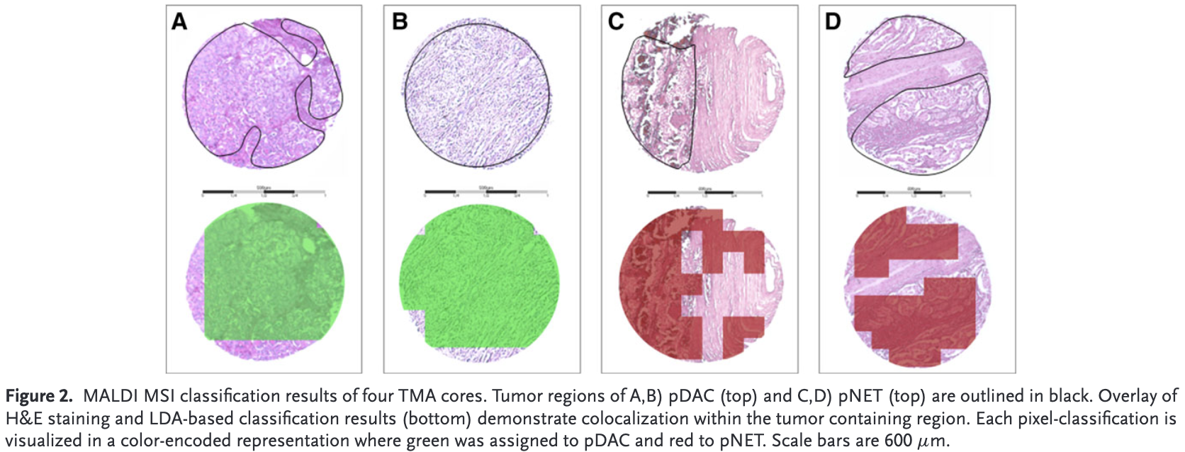
Purpose: To define proteomic differences between pancreatic ductal adenocarcinoma (pDAC) and pancreatic neuroendocrine tumor (pNET) by matrix-assisted laser desorption/ionization mass spectrometry imaging (MALDI MSI).
Experimental design: Ninety-three pDAC and 126 pNET individual tissues are assembled in tissue microarrays and analyzed by MALDI MSI. The cohort is separated in a training (52 pDAC and 83 pNET) and validation set (41 pDAC and 43 pNET). Subsequently, a linear discriminant analysis (LDA) model based on 46 peptide ions is performed on the training set and evaluated on the validation cohort. Additionally, two liver metastases and a whole slide of pDAC are analyzed by the same LDA algorithm.
Results: Classification of pDAC and pNET by the LDA model is correct in 95% (39/41) and 100% (43/43) of patients in the validation cohort, respectively. The two liver metastases and the whole slide of pDAC are also correctly classified in agreement with the histopathological diagnosis.
Conclusion and clinical relevance: In the present study, a large dataset of pDAC and pNET by MALDI MSI is investigated, a class prediction model that allowed separation of both entities with high accuracy is developed, and differential peptide peaks with potential diagnostic, prognostic, and predictive values are highlighted.
Link to full text: https://onlinelibrary.wiley.com/doi/10.1002/prca.201800046
Casadonte, R., Kriegsmann, M., Perren, A., Baretton, G., Deininger, S., Kriegsmann, K., Welsch, T., Pilarsky, C., & Kriegsmann, J. (2018). Development of a Class Prediction Model to Discriminate Pancreatic Ductal Adenocarcinoma from Pancreatic Neuroendocrine Tumor by MALDI Mass Spectrometry Imaging. In PROTEOMICS – Clinical Applications (Vol. 13, Issue 1, p. 1800046). Wiley. https://doi.org/10.1002/prca.201800046